An
echocardiogram is a device that sends high frequency sound waves (that cannot
be heard by the human ear) through a device called a transducer. The transducer
uses the sound waves that bounce back from the heart to create an image of
heart. It is much like an ultrasound used to view babies. This process of
obtaining these images is very quick, so by combining many images in a short
amount of time, a video can be created
that allows doctors to see how the heart is contracting and expanding.
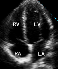
RV- right ventricle
LV- left ventricle
RA- right atrium
LA- left atrium
Transthoracic echocardiogram (TTE)
This is the most common echocardiogram preformed. The transducer is simply moved across the patient’s chest to obtain images.
STRESS echocardiogram
During this test, the transducer is moved across the patients test to obtain images when they are at a resting heart rate then again after they are at their target heart rate. The images are used to diagnose certain cardiac issues. The heart rate is normally raised by walking on a treadmill, however for patients that are unable to walk, a shot is given to raise the patient’s heart rate.
Trans esophageal echocardiogram (TEE)
Sometimes, a transducer cannot produce images that are clear enough for the doctor. During the TEE test, patients first have a sedative and anesthetic given to them and then they have a probe placed down their esophagus with a transducer like machine attached to the bottom. Since this is closer to the heart, without lungs and bones obstructing the sound waves, this device allows the doctor to get clearer images of the heart.
